There are four common methods of treatment used in the removal of ear wax. These are ear wax drops, ear syringing or irrigation, micro-suction ear wax removal and endoscopic ear wax removal.
Ear Wax Drops
There are many over the counter preparations available from any pharmacy to loosen, soften or dissolve ear wax. These will be either oil-based (olive oil) or water-based (sodium bicarbonate or saline). Water-based drops are considered to be more effective at dissolving ear wax than oil-based preparations. If a perforation has been diagnosed in the past, drops should be avoided, as they may cause issues with middle ear infections. The drops are applied at home and must be at room temperature to avoid the “caloric effect”, which is caused when the balance organ in the ears are out of sync. This is due to an inhibition in its function caused by the cooler air or water.
The application of ear wax drops can be messy and time-consuming. It requires the affected ear to be facing upwards when the drops are applied. Once applied, you must remain there for 5 to 10 minutes after which, you can sit up and wipe away any excess which will come out of the ear. This process needs to be applied 2/3 times per day for 10 to 14 days. Even after this procedure if the wax is impacted the use of drops alone may not be successful.
Ear Syringing (Irrigation)
Ear irrigation is normally performed by a GP practice nurse, a district nurse, and by some Audiologists. It is usually free on the NHS. Traditionally, a metal ear syringe was loaded with warm water, the metal tip placed into the ear canal. The water was then squirted into the ear canal and a kidney dish was held under the ear to catch the water and wax that was flushed out. The syringe would have to be regularly lubricated to allow a smooth level of pressure to be applied. The nurse would use his or her judgment as to how forcefully to syringe the water. Nowadays, for safety reasons, the metal ear syringe has been replaced by an ear irrigator pump with a jet tip. The pump has a variable, regulated pressure, but the process is essentially the same. Many people have had their ears syringed or irrigated many times without any issue arising.
Side Effects, Risks and Disadvantages
When it works it works well, however, ear syringing comes with some risks and disadvantages:
- Hard wax must be softened for up to two weeks before syringing.
- Syringing can push wax further into the ear if the angle of the jet is slightly off.
- May cause tinnitus.
- It may perforate the eardrum.
- An undiagnosed perforated eardrum may not be seen due to the amount of wax. This will cause water, bacteria, wax and dead skin cells will be flushed into the middle ear. This could potentially cause a painful infection.
- Is not recommended following ear surgery.
- Should not be performed when the eardrum has previously been perforated due to the risk of re-perforation.
- Ear syringing should under no circumstances be performed on individuals with a known perforation, cleft palate, foreign object in the ear canal or a mastoid cavity following a mastoidectomy.
- Many surgeries are withdrawing their ear syringing service and are referring all patients to the NHS ENT clinic. The
- NHS ENT clinic may have a long waiting list.
Micro-Suction Ear Wax Removal
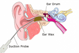
Micro-suction is normally performed in hospital outpatient clinics by ENT doctors and nurses when other methods have not been unsuccessful. The major benefit of micro-suction is that the wax is removed safely under direct vision at all times. In a hospital, the procedure is undertaken using an ENT binocular microscope to clearly visualize the wax. A sterile suction probe connected to a gentle suction machine is then used. Other ENT instruments can also be used. Unlike irrigation or syringing micro-suction is a “dry” procedure and does not require the use of water. This significantly reduces the risk of damage to
How Does it Work?
The use of micro-suction is quicker and safer and does not require weeks of ear drops. Micro-suction in ENT departments is performed using large expensive ENT binocular operating microscopes. Due to waiting
Side Effects and Risks
Due to the suction machine, this process can occasionally be noisy due to the suction machine. Sometimes flaps of dry skin vibrate causing ‘clareneting’. Subsequently like ear syringing people report tinnitus or increased tinnitus if they already experience it before micro-suction. There is also a slight chance of bleeding due to grazing or scratching
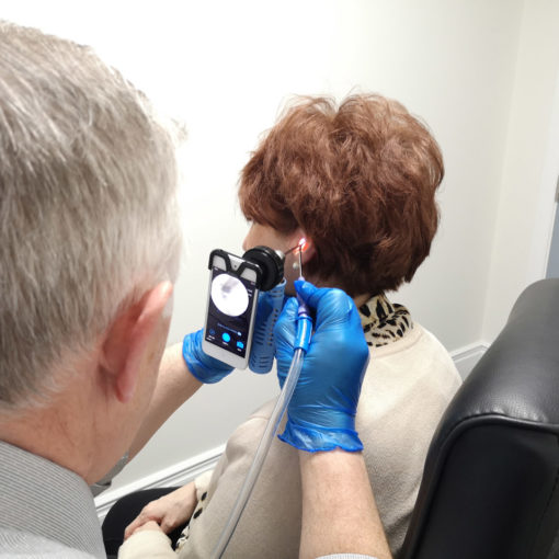
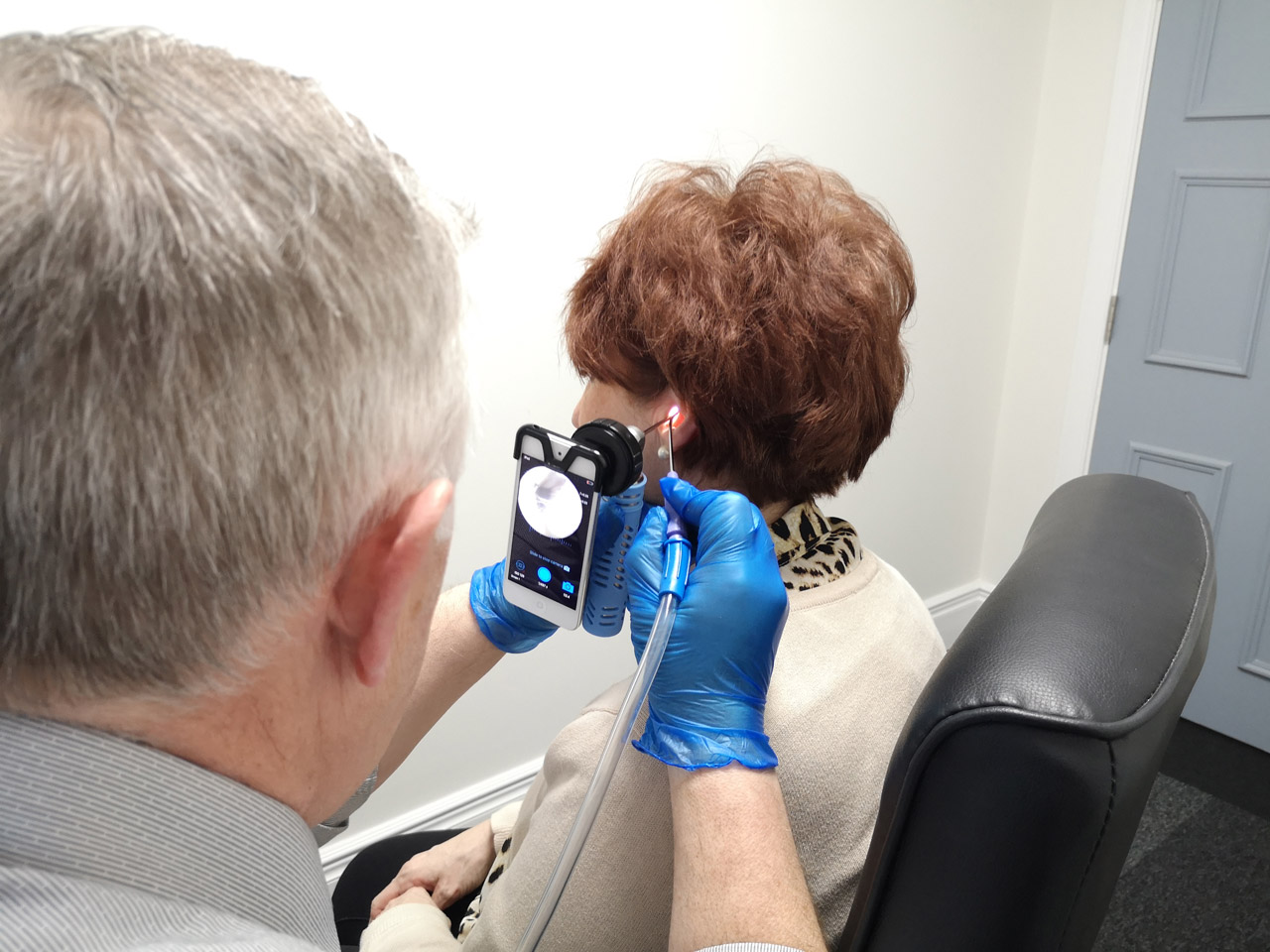
Endoscopic Ear Wax Removal

An alternative to micro-suction is endoscopic ear wax removal, it is quicker and easier to perform than micro-suction. To perform endoscopic ear wax removal an
A 2010 Health Technology Assessment by AJ Clegg et. al. concluded that endoscopic ear wax removal is superior to microscopic wax removal – microscopic wax removal uses an operating microscope or surgical loupes to get a magnified view of the ear canal and ear drum – a speculum (small funnel) must be inserted into the ear canal in order to provide a view, and the speculum takes up some of the space within the ear canal, further restricting the practitioner’s view. Furthermore, the speculum can sometimes cause minor trauma to the skin of the ear canal if the ear canal is very narrow.
How Does it Work?
In contrast, endoscopic ear wax removal uses a fine 2.8mm endoscope inserted part way into the ear canal. The endoscope is a rod containing fiber-optics to relay light from a light source to illuminate the canal and a solid lens (or series of lenses) to relay the view from inside the canal to the outside. The endoscope can be either viewed directly, or imaging equipment can be attached to provide better magnification
Side Effects and Risks
As with micro-suction, endoscopic ear wax removal can be noisy. There is a very small risk, as with all other wax removal treatments, that some people may report tinnitus or increased tinnitus if they already experienced it pre- ear wax removal. There is a small chance of slight bleeding due to grazing and scratching the ear canal with the suction probe. However, compared to micro-suction, this risk is further reduced because there is more o

